MYOMS
• Abnormal bleeding
• Painful intercourse
• Abdominal swelling, constipation
• Frequent need to urinate etc.
If you have similar complaints, the cause may be myoma.
MYOMA
Myomas, one of the most common gynecological diseases today, are benign tumors that occur as a result of abnormal smooth muscle proliferation in the uterus. Myomas rarely occur during adolescence and young adulthood. It is seen in 20% of women of reproductive age and is the most common reason for hysterectomy surgery. Myomas, which are often formed between the ages of 30 and 40, tend to shrink with menopause.
Myomas that grow after menopause may need to be monitored for malignant transformation. The probability of myomas becoming malignant (cancerous) is 0.1%. Rapid growth of myomas is an indication for surgery. Although the main cause of myoma formation is estrogen hormone, it is known that familial predisposition also plays a role. Being overweight increases the tendency to form myomas. They can grow in size and cause pain during pregnancy and, large size subserous fibroids which compress the uterine cavity, can cause infertility, miscarriage, recurrent pregnancy loss, and preterm delivery.
SYMPTOMS OF MYOMAS
Fibroids, which can occur at any time in the life of many women, often go undetected. In cases where it does not cause symptoms, the person can live with myoma for life. However, some patients may experience symptoms such as abnormal bleeding, disturbingly painful and bleeding menstrual periods, back pain and painful sexual intercourse. Myomas that reach large sizes can cause abdominal swelling, pain, indigestion, constipation and gas complaints. It can make one feel the need to urinate frequently by pressing on the bladder. Fibroids that tend to grow, overgrow, or cause symptoms can even lead to infertility. Stem myomas inside the cavity may, in rare cases, come out of the uterine cavity and cause post-intercourse bleeding and discharge with a foul odor due to infection.
TYPES OF MYOMAS
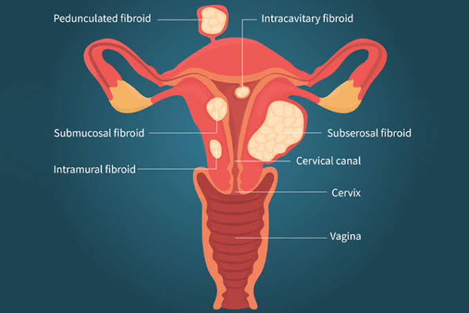
Myomas are classified according to their location on the uterine wall: • Submucous myoma: Those located on the inner wall of the uterus • Intramural myoma: Those embedded in the muscle layer of the uterus • Subserous myoma: Those that have grown outside the uterus • Stem and subserous myoma: Those that are connected to the outside of the uterus with a stalk. Myoma may show symptoms depending on its location, number and size.
DIAGNOSIS METHODS
Myomas usually appear as a result of routine gynecological examinations. The clinical diagnosis of fibroids is made by obstetrical examination and ultrasonography. The size and location of the myoma is determined by using high-resolution ultrasound. In some cases, to understand/determine the inner wall of the uterus and the proportion of the myoma inside the uterus versush the muscle layer, a method called saline infusion sonography known as "aqueous ultrasonography" can be applied. In this method, fluid is injected into the uterus with a thin cannula, and the inner layer of the uterus is visualized with ultrasonography. Thus, the location and extension of the fibroid is observed. In multiple myomas, one of the imaging methods used to map is medicated MRI. With MRI imaging, detailed information on soft tissues can be obtained.
MYOMAS TREATMENT
Most myomas, which are not causing any symptoms or complaints are detected incidentally during gynecological examination. In such myomas, the myoma is usually monitored periodically without any treatment. Myoma treatment can be done with medication. Estrogen and progesterone hormone production is blocked by drugs defined as GnRH (Gonadotropin-releasing hormone). Thanks to this treatment, which causes the person to enter menopause temporarily, menstrual bleeding stops and myomas shrink. One of the treatment methods that does not require surgery is radiofrequency (RF) ablation. Radiofrequency energy is administered to the myoma by entering it with a special needle. This type of treatment, which causes an increase in temperature within the myoma, requires anesthesia. In embolization treatment, which is one of the minimally invasive procedures, the blood flow to the small vascular structures feeding the myoma is interrupted. Myomas that cannot be nourished will shrink over time. Surgery is preferred if the fibroid is enlarged or if it is a problem for the patient. The decision for surgery is made based on the patient's age, complaints, number and location of myomas, and whether the patient has children, and the scope of the surgery is determined. The surgical method used in the treatment of myoma is defined as myomectomy. Myomectomy for myomas suitable for closed surgery is performed laparoscopically and the myomas in the uterus are surgically removed. After myomectomy, reproductive potential increases, but some problems such as myoma reformation, bleeding, hematoma, and adhesion may occur. The only treatment method with proven efficacy in the treatment of fibroids is hysterectomy. In the hysterectomy procedure, the uterus is completely removed. In appropriate cases, closed surgery may be preferred. In patients of reproductive age, the patient does not enter menopause unless eggs are taken as an additional procedure.
 Language
  Türkçe
 English